)


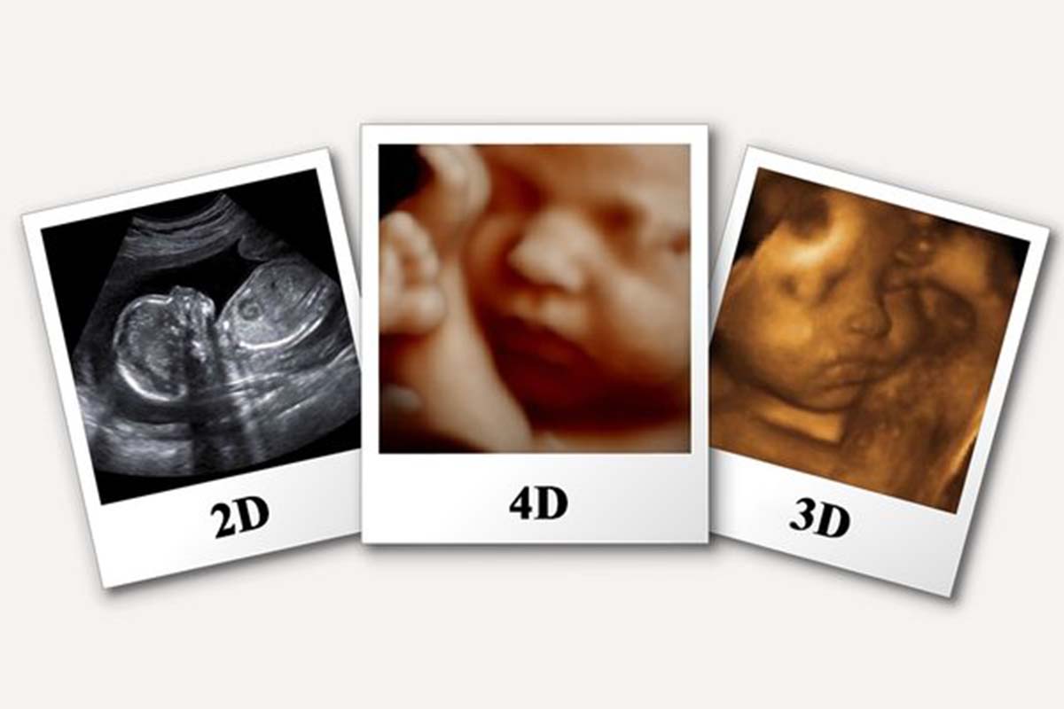
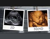
3D and 4D sonography are advanced imaging techniques that provide detailed visualizations of the developing fetus during pregnancy. Unlike traditional 2D ultrasound, these technologies create three-dimensional images and dynamic video sequences that enhance the understanding of fetal anatomy and well-being.

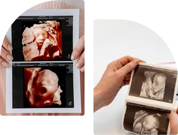
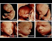
3D sonography captures multiple two-dimensional images of the fetus from various angles and combines them to create a three-dimensional image. This technology allows healthcare providers and expectant parents to see more detailed features of the fetus, including facial characteristics and limb development.
4D sonography takes the concept of 3D imaging a step further by adding the dimension of time. It produces real-time moving images of the fetus, allowing parents to witness their baby’s movements, such as kicking, yawning, and even smiling. This adds an emotional connection to the prenatal experience.
.jpg)
 Final.jpg)
3D sonography provides clearer, more detailed images of the fetus, helping identify physical abnormalities or congenital defects earlier in pregnancy.
4D sonography offers live video of the fetus, allowing parents to see their baby's movements, which can provide reassurance about the baby's health.
Expecting parents can see their baby's features and movements, fostering a deeper emotional bond before birth.
Many parents choose to create keepsakes, such as photos and videos, from 3D/4D scans to cherish the memories of their pregnancy journey.
The detailed imaging helps healthcare providers assess fetal growth and development, leading to better management of potential complications.
If abnormalities are detected, early intervention can be planned, improving outcomes for both mother and baby.
3D and 4D sonography are safe, non-invasive procedures that do not involve any radiation, making them a preferred choice for monitoring fetal health.
Proactively revolutionize granular customer service after pandemic internal or "organic" sources impact proactive human
Used as part of routine prenatal screenings to monitor fetal development and well-being.
Helps in identifying physical defects or abnormalities in the fetus, such as cleft lip, heart defects, or spinal abnormalities.
Provides insight into the baby's position and helps in planning for labor and delivery.
3D and 4D sonography have revolutionized prenatal care by providing clearer images and real-time visuals of the developing fetus. These advanced imaging techniques enhance emotional bonding, improve diagnostic capabilities, and support expectant parents throughout their pregnancy journey. If you're looking to experience the magic of seeing your baby before birth, 3D/4D sonography is an invaluable option to consider.